
Understanding Natural Killer (NK) Cells in Cancer Biology
Natural Killer (NK) cells are large granular lymphocytes comprising 5% to 15% of human peripheral blood mononuclear cells (PBMCs). Classified as key effectors of the innate immune system, NK cells possess a distinct immunological advantage: they can identify and eliminate transformed malignant cells rapidly without prior antigen sensitization or clonal expansion.
Unlike cytotoxic T lymphocytes (CTLs), which depend entirely on the presentation of tumor antigens bound to Major Histocompatibility Complex (MHC Class I) molecules, NK cells function via the seminal "missing-self" hypothesis. Many aggressive cancers strategically downregulate or lose MHC Class I surface expression to hide from cytotoxic T cells. NK cells specifically detect this absence of MHC Class I molecules, recognizing the cell as abnormal and triggering targeted cytolysis.
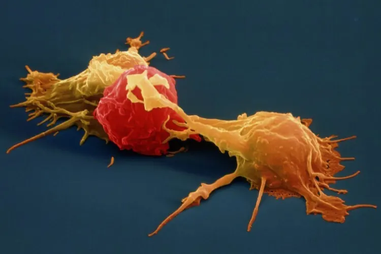 Figure: Activated Natural Killer (NK) cell binding to malignant target cell and releasing cytolytic granules.
Dual Receptor Balance & Cytolytic Killing Mechanisms
NK cell activation is tightly governed by a delicate equilibrium between activating and inhibitory cell-surface receptors:
Receptor-Mediated Target Recognition
Activating receptors such as NKG2D, DNAM-1, and Natural Cytotoxicity Receptors (NKp30, NKp44, NKp46) recognize stress-induced ligands (MIC-A, MIC-B, ULBPs) overexpressed on tumor membranes, tipping the intracellular balance toward activation when inhibitory Killer-cell Immunoglobulin-like Receptors (KIRs) find no normal MHC Class I ligands.
Perforin & Granzyme B Granule Exocytosis
Upon immunological synapse formation, NK cells release pre-formed cytotoxic granules containing perforin, which polymerizes to create pore channels in the malignant cell membrane, permitting granzyme B entry to initiate rapid caspase-dependent apoptosis.
Death Receptor Pathway Activation
Activated NK cells express tumor necrosis factor-related apoptosis-inducing ligand (TRAIL) and Fas Ligand (FasL), engaging death receptors (DR4/DR5 and Fas/CD95) on tumor cells to induce extrinsic apoptotic cascades.
Antibody-Dependent Cellular Cytotoxicity (ADCC)
Via high-affinity CD16 (FcγRIIIa) surface receptors, NK cells bind the constant region of therapeutic monoclonal antibodies (such as rituximab, trastuzumab, or cetuximab), producing potent synergistic target killing.
The Clinical Protocol: Autologous Immune Enhancement Therapy (AIET)
Our clinical partners utilize standardized, multi-step ex-vivo culture protocols to overcome the quantitative and qualitative deficits common in cancer patients' endogenous NK cells:
Step 1: Blood Collection
50–80 mL of peripheral blood or mild leukapheresis is drawn to harvest mononuclear cells (PBMCs) under sterile outpatient conditions.
Step 2: cGMP Expansion
Cells are cultured in specialized cleanrooms with Interleukin-2 (IL-2), Interleukin-15 (IL-15), and feeder layers for 14–21 days, expanding NK cells up to 500- to 1,000-fold.
Step 3: Quality Infusion
After passing flow cytometry purity (>80% CD3- CD56+), endotoxin, and viability tests (>90%), highly activated NK cells are re-infused intravenously over 60 minutes.
Malignancies Under Investigational Evaluation
NK cell adoptive immunotherapy is explored across an array of solid tumors and refractory hematologic malignancies where immune escape mechanisms are pronounced:
Solid Tumors
- Colorectal & Gastrointestinal Malignancies: Eradicating circulating tumor cells
- Non-Small Cell Lung Cancer (NSCLC): Adjuvant maintenance post-chemoradiotherapy
- Ovarian & Breast Cancers: Overcoming MHC-I downregulation and refractory lesions
- Renal Cell & Hepatocellular Carcinomas: Intercepting micrometastatic dissemination
Hematologic Malignancies
- Acute Myeloid Leukemia (AML): Targeting residual leukemic blasts post-consolidation
- Multiple Myeloma: Targeting marrow niche clonal plasma cells
- Non-Hodgkin Lymphomas: Synergized with anti-CD20 monoclonal antibody therapy
- Minimal Residual Disease (MRD): Lowering relapse probability
Clinical Candidacy & Pre-Screening Requirements
Candidates are evaluated via multi-specialty oncology boards to determine individual physiological readiness:
✓ Potential Candidates
- Histopathologically confirmed solid or hematologic malignancy
- Adequate general performance: ECOG status 0 to 2
- Adequate absolute lymphocyte count (ALC ≥ 800/μL) and platelet count (≥ 80,000/μL)
- Sufficient interval post-chemotherapy (minimum 2–3 weeks washout period)
- Realistic clinical expectations focusing on immune surveillance and quality of life
✗ Exclusion Factors
- Severe terminal end-organ failure (severe hepatic cirrhosis, advanced renal shutdown)
- Uncontrolled systemic bacteremia, active fungal infections, or sepsis
- Severe active autoimmune disease on aggressive immunosuppression
- Current concurrent high-dose corticosteroid treatment suppressing NK cytotoxicity
- Profound cytopenias unresponsive to hematopoietic support
Cost Comparison: India vs Global Centers
Through India's advanced biotechnology infrastructure, international patients access international-grade cleanroom cellular expansion at transparent price points:
| Country / Destination | Average Treatment Package | Typical Turnaround | Accreditation & Standards |
|---|---|---|---|
| India (Our Partner Centers) | $4,500 – $7,500 USD | 2 – 3 Weeks | JCI / NABH & cGMP Certified |
| Japan | $28,000 – $48,000 USD | 3 – 5 Weeks | PMDA Regenerative Framework |
| Germany / UK | $32,000 – $55,000 USD | 1 – 3 Months | EU Advanced Therapy (ATMP) |
| United States | $40,000 – $80,000 USD | 2 – 4 Months | Clinical Trial Protocols Only |
What Is Included in the India NK Cell Package?
Blood isolation / leukapheresis, 14–21 day cGMP expansion with clinical cytokines, batch release sterility & endotoxin assays, flow cytometry characterization (CD3- CD56+), multi-session intravenous re-infusion under clinical telemetry, oncologic review, airport transfers, and personal medical coordination.
Peer-Reviewed Scientific Literature
Our protocols reflect ongoing scientific advancements reported in leading peer-reviewed oncology and immunology journals:
- Vivier, E. et al. (2012): "Innate or adaptive immunity? The example of natural killer cells." Science, 331(6013): 44-49.
- Shimasaki, N., Jain, A., & Campana, D. (2020): "NK cells for cancer immunotherapy." Nature Reviews Drug Discovery, 19(3): 200-218.
- Romee, R. et al. (2016): "Cytokine-induced memory-like natural killer cells exhibit enhanced function against myeloid leukemia." Science Translational Medicine, 8(357): 357ra123.
- Ishikawa, E. et al. (2007): "Autologous natural killer cell therapy for glioblastoma multiforme." Journal of Neuro-Oncology, 83(2): 185-194.