
Understanding Dendritic Cells in Tumor Immunology
Dendritic cells (DCs) are the professional antigen-presenting cells (APCs) of the human immune system. Discovered by Nobel laureate Ralph Steinman, DCs serve as the critical immunological bridge between the innate and adaptive immune responses.
In healthy biological states, dendritic cells continuously capture foreign peptides, process them into antigenic epitopes, migrate to regional draining lymph nodes, and present these antigens via Major Histocompatibility Complex (MHC Class I and Class II) molecules to naive CD8+ cytotoxic T lymphocytes and CD4+ helper T cells. This triggers a clonal expansion of targeted cytotoxic effector cells.
In malignant diseases, however, tumors create an immunosuppressive microenvironment. Malignant cells downregulate MHC expression, secrete inhibitory cytokines (such as TGF-β, VEGF, and IL-10), and recruit regulatory T cells (Tregs) and myeloid-derived suppressor cells (MDSCs). This renders endogenous dendritic cells dysfunctional or immature, preventing the immune system from recognizing and eliminating malignant cells.
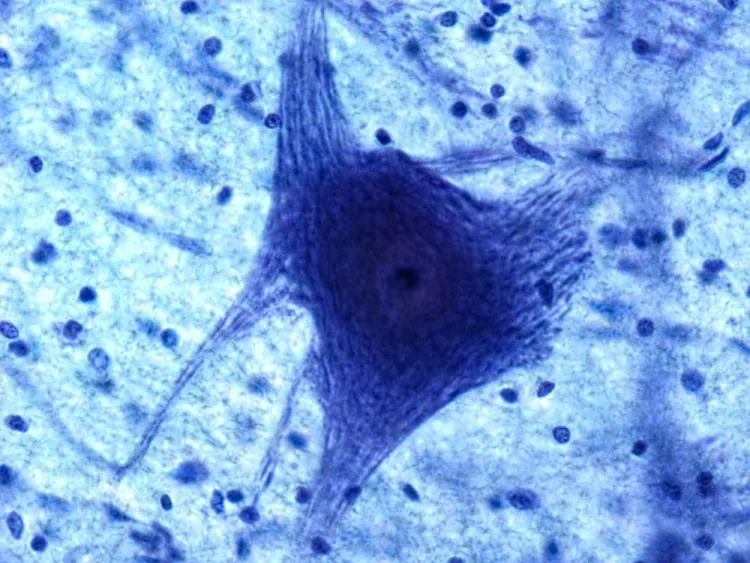 Figure: Autologous dendritic cell maturation, antigen pulsing, and cytotoxic T-lymphocyte priming in clinical oncology.
Mechanism: How Dendritic Cell Therapy Operates
Autologous dendritic cell therapy bypasses tumor-mediated immune paralysis by culturing, maturing, and arming the patient's own immune cells outside the hostile tumor environment:
Monocyte Isolation via Peripheral Apheresis
Mononuclear cells (PBMCs) are separated from the patient's peripheral blood via gentle leukapheresis. CD14+ monocytes are isolated under certified ISO Class 5 cleanroom conditions.
Ex-Vivo Differentiation & Cytokine Maturation
Monocytes are cultured in clinical-grade media supplemented with GM-CSF (Granulocyte-Macrophage Colony-Stimulating Factor) and Interleukin-4 (IL-4) to differentiate into immature dendritic cells, followed by maturation factors (TNF-α, IL-1β, PGE2).
Tumor Antigen Loading & Epitope Pulsing
Mature dendritic cells are pulsed with patient-derived tumor lysates, specific synthetic peptide antigens (e.g., MUC1, HER2, WT1, CEA, or NY-ESO-1), or neoantigens, programming them to recognize the patient's unique malignant profile.
Re-infusion & Systemic T-Cell Priming
Activated, antigen-loaded dendritic cells are re-administered intradermally near lymphatic basins or intravenously. Once inside the lymphatic circulation, they present tumor antigens to naive T cells, generating systemic, durable anti-tumor memory.
Clinical Applications Under Investigation
Dendritic cell protocols are evaluated as supportive adjuvants in various solid tumors and hematologic malignancies, including:
Epithelial & Solid Malignancies
- Non-Small Cell Lung Cancer (NSCLC): Adjuvant surveillance post-chemo/radiotherapy
- Colorectal & Gastric Cancers: CEA/MUC1 targeted cellular immunization
- Breast Cancer: HER2-positive and refractory triple-negative phenotypes
- Prostate Cancer: Investigated in metastatic castration-resistant models
High-Risk & Refractory Tumors
- Renal Cell Carcinoma (RCC): Highly immunogenic tumor profiling
- Cutaneous Melanoma: Early and late-stage antigen-primed surveillance
- Ovarian Carcinoma: Post-debulking minimal residual disease control
- Glioblastoma Multiforme (GBM): Autologous lysate-pulsed protocols
Standardized Administration Routes
Our partner NABH and JCI-accredited cancer centers utilize evidence-aligned delivery methods based on tumor topography and stage:
| Administration Route | Clinical Rationale | Procedure Details |
|---|---|---|
| Intradermal Injection (Proximal Lymph Nodes) | Provides direct access to afferent lymphatic vessels, maximizing presentation efficiency to nodal T-cell zones. | Microneedle injection into skin folds adjacent to axillary, inguinal, or cervical lymph basins. |
| Intravenous (IV) Infusion | Facilitates widespread systemic circulation to prime splenic and circulating T-cell populations. | Delivered in sterile saline over 45–60 minutes under continuous vital sign monitoring. |
| Intratumoral Infiltration (Selective) | Directly targets primary tumor beds or accessible metastatic nodes to reverse local microenvironmental paralysis. | Conducted under high-resolution ultrasound or CT guidance by interventional radiologists. |
Patient Candidacy & Comprehensive Screening
Every patient undergoes multi-disciplinary tumor board evaluation to confirm safety, immune responsiveness, and realistic clinical parameters:
✓ Potential Candidates
- Histologically confirmed solid tumor or lymphoproliferative disorder
- Adequate functional performance: ECOG status 0 to 2
- Preserved bone marrow function (Absolute Neutrophil Count ≥ 1,500/μL, Platelets ≥ 100,000/μL)
- Adequate renal (creatinine ≤ 1.5x ULN) and hepatic profiles
- Patients seeking supportive cellular therapy to complement or follow standard oncologic care
✗ Exclusion Factors
- Severe terminal organ decompensation (NYHA Class IV heart failure, severe respiratory failure)
- Active uncontrolled systemic sepsis or bacterial/fungal infection
- Severe active autoimmune disease requiring heavy systemic immunosuppression
- Severe coagulopathy preventing safe peripheral venipuncture or leukapheresis
- Concurrent high-dose immunosuppressive therapy (e.g., prednisone > 20mg/day)
Cost Breakdown: India vs International Centers
Receiving specialized cell-based cancer support in accredited Indian hospitals delivers state-of-the-art cGMP laboratory processing at substantial cost efficiency:
| Country / Region | Average Treatment Cost | Typical Waiting Period | Facility Accreditation |
|---|---|---|---|
| India (Our Partner Centers) | $4,500 – $7,500 USD | 1 – 2 Weeks | JCI / NABH & cGMP Cleanroom |
| United States | $45,000 – $95,000 USD | 2 – 4 Months | FDA Cleared / Trial Only |
| Germany / Switzerland | $35,000 – $65,000 USD | 1 – 3 Months | EU GMP Certified |
| Japan | $25,000 – $42,000 USD | 3 – 6 Weeks | PMDA Regulated |
What Is Included in the India Immunotherapy Package?
Mononuclear cell leukapheresis, cGMP cleanroom maturation and antigen loading, safety release testing (mycoplasma, endotoxin, flow cytometry purity > 85%), 4 to 6 scheduled dose administrations, medical oncologist consultations, pre-treatment laboratory panels, local airport concierge transfers, and multi-lingual coordinator support.
Peer-Reviewed Scientific Literature
Our medical perspective is grounded in published clinical trials evaluating dendritic cell vaccines in global oncology literature:
- Kantoff, P. W. et al. (2010): "Sipuleucel-T Immunotherapy for Castration-Resistant Prostate Cancer." New England Journal of Medicine, 363(5): 411-422.
- Anguille, S. et al. (2014): "Clinical use of dendritic cells for cancer therapy." The Lancet Oncology, 15(7): e257-e267.
- Santos, P. M., & Butterfield, L. H. (2018): "Dendritic Cell-Based Cancer Vaccines." The Journal of Immunology, 200(2): 443-449.
- Garg, A. D. et al. (2017): "Trial watch: Dendritic cell-based anticancer immunotherapy." OncoImmunology, 6(7): e1328341.